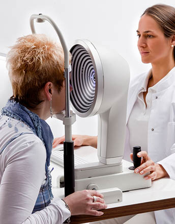
The Oculus 5M is a incredibly powerful tool for diagnosis and education. Dr. Brimer has actually worked with the Oculus team in Germany for several years helping to design the current Oculus platform and the Crystal Tear Report. This version of the 5M is successful globally and we are so thankful to have been able to empower doctors around the world to better help their patients. Dr. Brimer knows this device better than anyone and can use it to let you SEE elements of your ocular surface that are impossible to see on a regular exam with a slit lamp.
Below are examples of what we can do and see with the Oculus 5M:
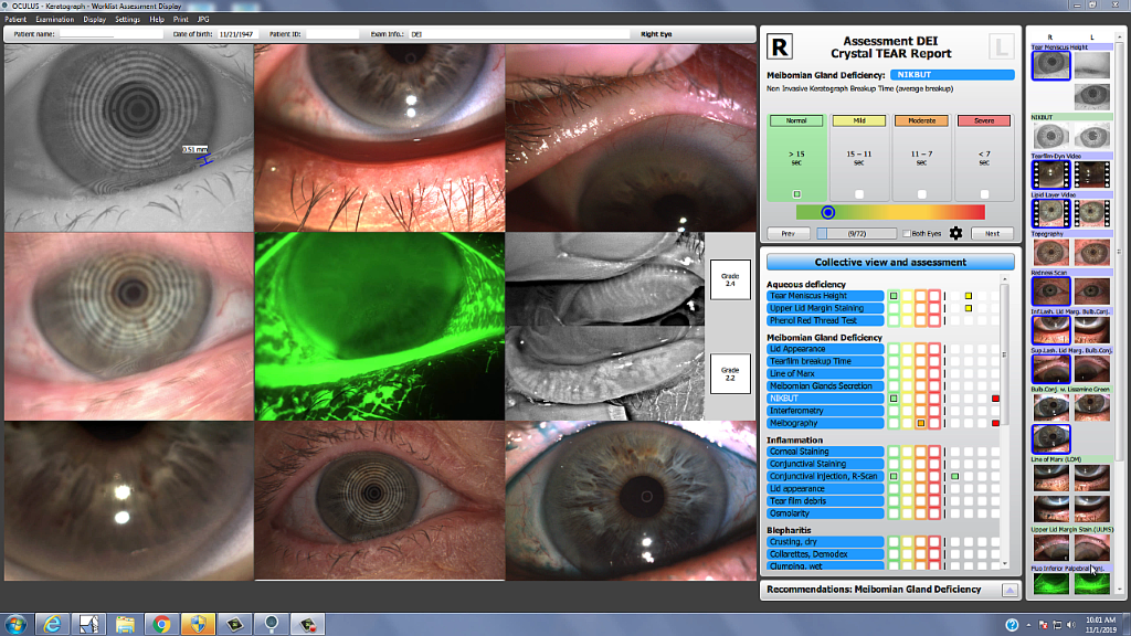
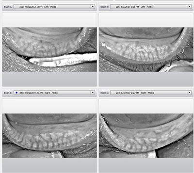
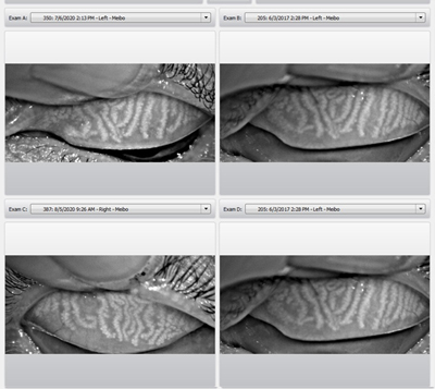
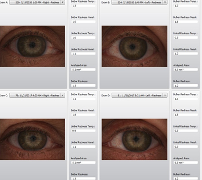
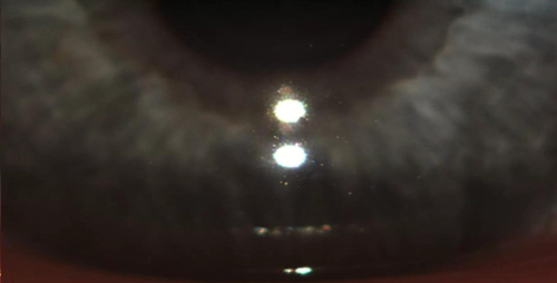
At every exam we will take a collage of images so you know your current status and can see the progress that has been made.
We are able to video the amount of oil being excreted into the tear film by your oil glands.
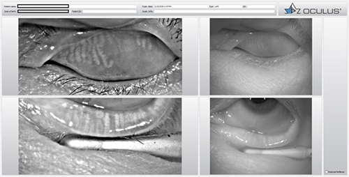
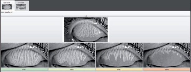
We can actually image and grade your oil glands.
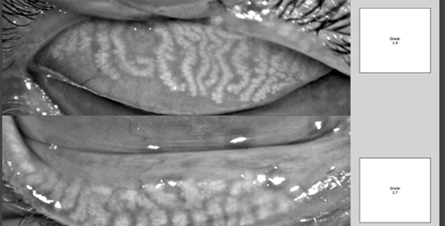
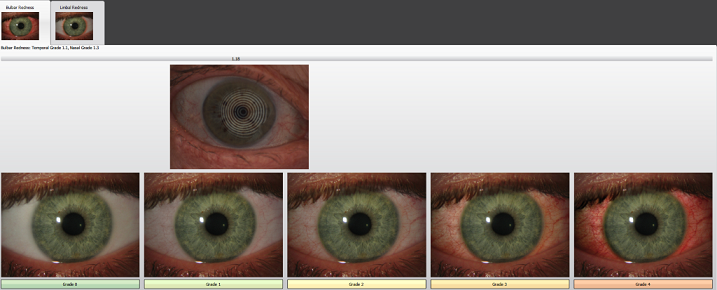
Show you how it compares to normal.
And how it remains stable or changes over time.
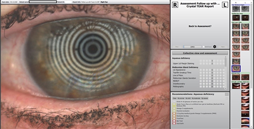
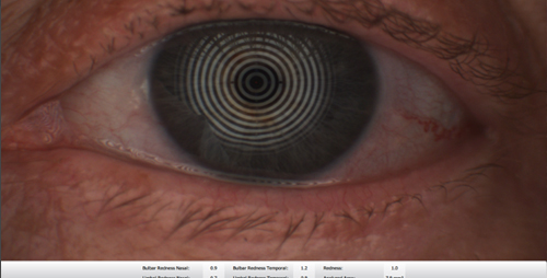
We can measure the tear volume for future comparison.


We can scale the redness in your eyes for future comparison.
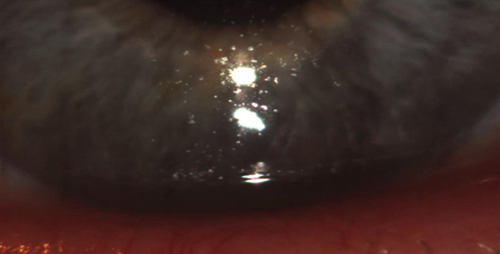
We can video the debris in your tear film.
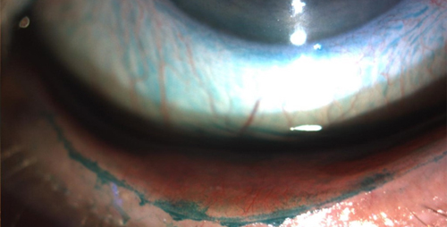
We can use special dyes to show damaged cells.
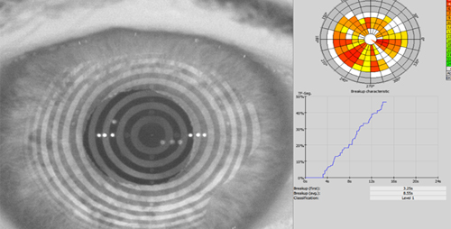
We can time and video your tear film actually evaporating.

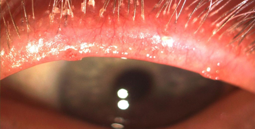
We can take magnified images of the lid margin to show lid bacteria and mites.
After your initial exam, you will receive your Crystal Tear Report. This will show your primary and secondary issues as well as your results for every test we did, in pie graph form. The results are color coded, with the stop light colors of green being normal and red, severe. You will see which treatments are specific to which problem, a definition of every test we did, and a list of all the recommended treatments.

710 Military Cutoff Rd Suite 130, Wilmington, NC 28405
(910) 447-2020 | info@dryeyeequaton.com
Follow us on:
Crafted with Love: DigiCorns
